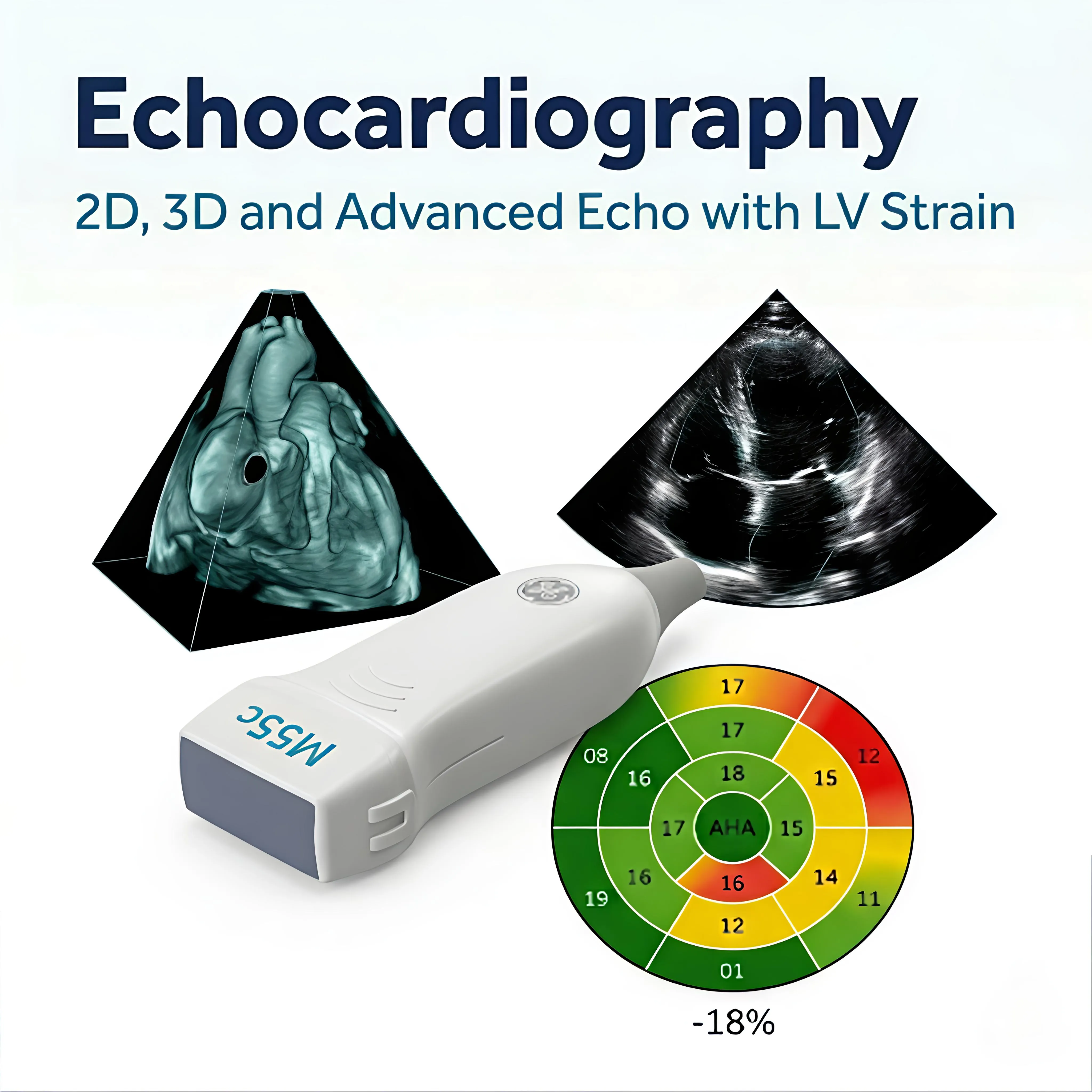
Echocardiography: A Detailed Ultrasound of Your Heart
2D, 3D, and Advanced Echo With GLS Understanding Your Heart's
Structure and Function
Echocardiography, an ultrasound
of the heart, is one of the most useful and versatile investigations in cardiology.
It is painless, uses no radiation, takes 20-45 minutes, and gives a
cardiologist a detailed real-time picture of how the heart looks and how it is
working. At Apollo CVHF, we offer the full range of echocardiographic
techniques, from standard 2D echo through to advanced 3D imaging and global
longitudinal strain (GLS) analysis.
What Is Echocardiography?
Echocardiography uses
high-frequency sound waves to produce images of the heart's chambers, valves,
and surrounding structures. A probe placed on the chest wall sends and receives
sound waves that bounce off the heart, producing a moving image of the heart in
real time.
Unlike X-rays or CT scans,
echocardiography uses no ionising radiation. It is safe to repeat as often as
needed for monitoring purposes.
Types of Echo Available at Apollo CVHF
•
2D Echocardiography : the
standard echo. Produces two-dimensional images of the heart's chambers and
valves, measures the size and function of each chamber, and assesses valve
opening and closing. The starting point for most cardiac assessments.
•
3D Echocardiography produces three-dimensional reconstructed images of the heart's structures.
Particularly valuable for detailed assessment of heart valves before surgical
or catheter-based intervention.
•
Echo with GLS (Global
Longitudinal Strain) an advanced technique that analyses subtle changes in
how the heart muscle contracts, detecting early dysfunction before it becomes
visible on standard 2D measurements. Particularly useful in patients on
chemotherapy, patients with diabetes, and those with early heart muscle
disease.
What Does Echo Assess?
•
The size and function of
the left and right ventricles
•
How well the heart is
pumping (ejection fraction)
•
The structure and function
of all four heart valves
•
The pericardium (the sac
surrounding the heart)
•
Blood flow patterns through
the chambers
•
Early detection of heart
muscle disease
What Should I Expect?
You lie on a couch and the
sonographer applies gel to your chest, then moves a handheld probe across the
chest wall to capture images from different angles. The procedure is entirely
painless. Results are reviewed by a cardiologist and discussed with you.