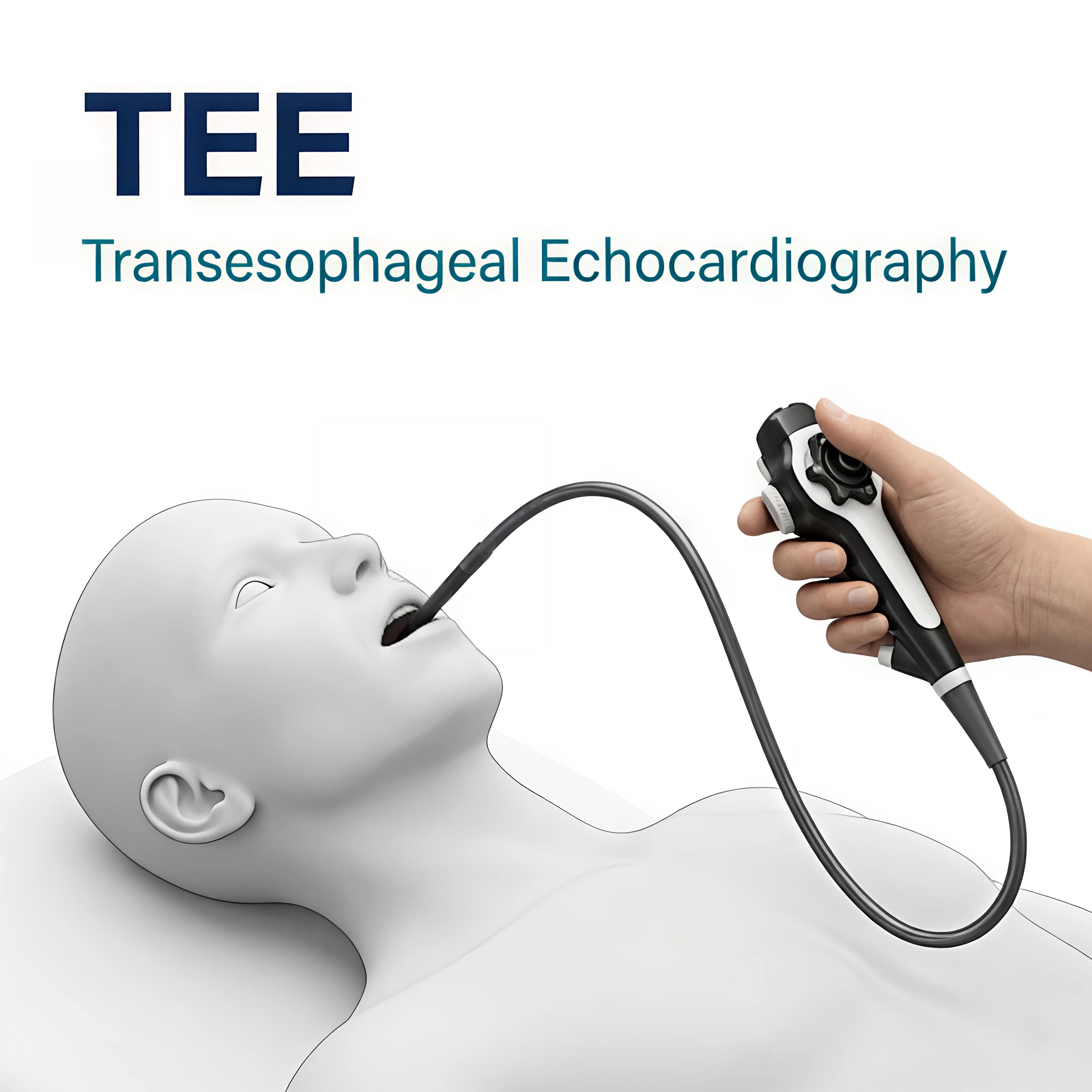
TOE Transesophageal Echocardiography
When Standard Echo Is Not Enough, Detailed Heart Imaging From
Inside
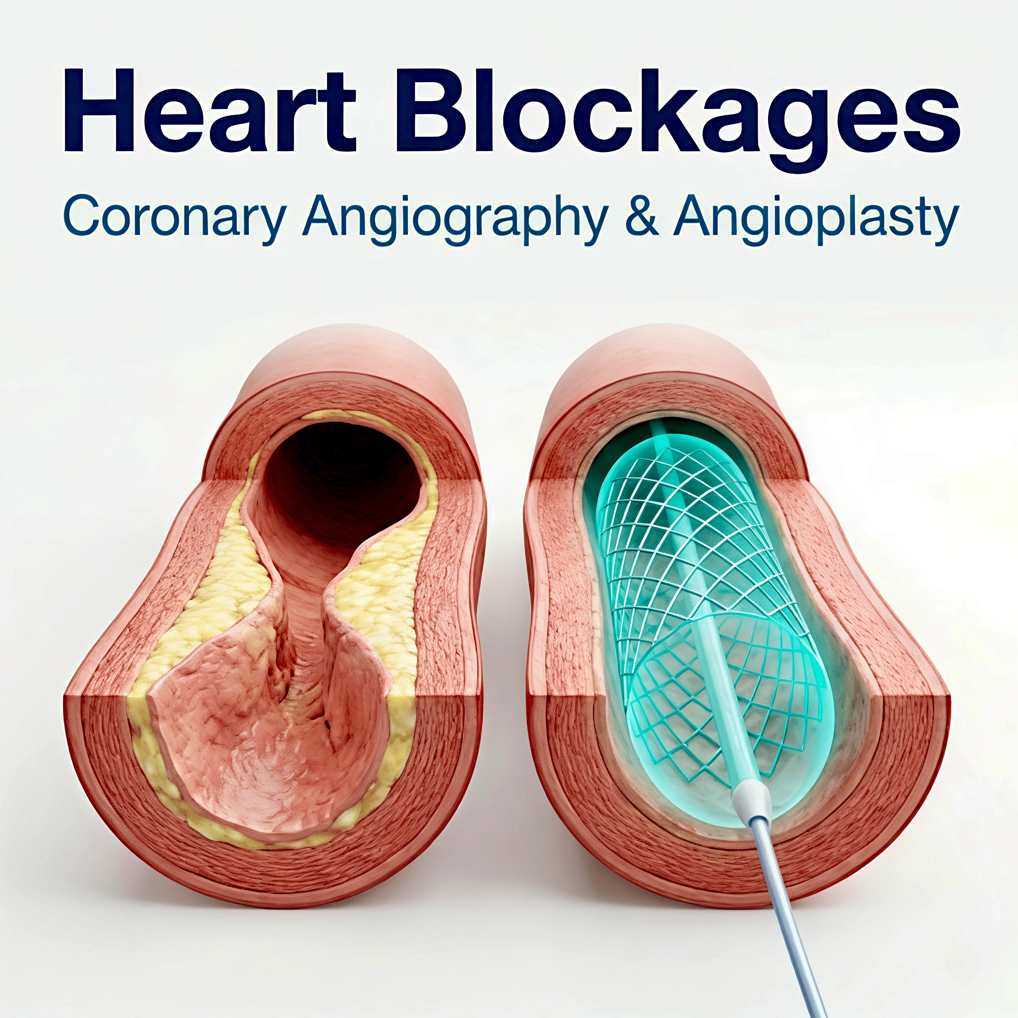
Standard echocardiography produces
excellent images for most patients, but in some situations, a clearer view is
needed. Transesophageal echocardiography (TOE) places the ultrasound probe
inside the oesophagus, the food pipe that runs immediately behind the heart, allowing much more detailed images of the heart's structures than can be
obtained from outside the chest wall.
When Is TOE Used?
•
Assessment of heart valves
before surgical or catheter-based intervention , particularly the mitral valve
•
Detection of blood clots
inside the heart's chambers, especially in patients with atrial fibrillation, before cardioversion or ablation
•
Detailed evaluation of the
aorta (the main artery from the heart)
•
Assessment of congenital
heart defects where more detail is needed
•
During cardiac procedures
such as TAVI or ASD closure, to guide device placement in real time
•
Investigation of a potential
source of stroke or embolism
What Does the Procedure Involve?
The procedure is carried out under
conscious sedation, you are relaxed and comfortable, but not under general
anaesthesia. The throat is sprayed with local anaesthetic. A flexible probe,
slightly thicker than a standard gastroscopy scope, is gently passed through
the mouth and into the oesophagus. The test typically takes 20-40 minutes.
You will need to fast for at least
4 hours beforehand. You will not be able to drive immediately after due to the
sedation, so please arrange for someone to accompany you.
|
TOE
is performed by experienced cardiologists and echocardiographers. The
procedure is well tolerated by most patients and is considerably less
uncomfortable than most people anticipate. |
After the Procedure
You will rest for 30-60 minutes
after the sedation before being discharged. Your throat may feel slightly sore
for the rest of the day. Results are typically discussed with you before you
leave or at a follow-up appointment shortly afterwards.