
Medically authored and audited by Dr. Kulin Sheth, Interventional Cardiologist
The choice between angioplasty (PCI) and bypass surgery (CABG) is rarely a matter of one being universally "better" than the other. They are distinct strategies for restoring blood flow to the heart muscle, and the optimal choice depends entirely on a patient's unique cardiovascular profile, anatomy, and biological risk factors.
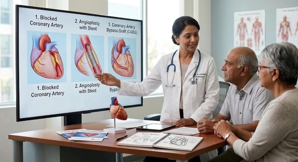
Generally, angioplasty with drug-eluting stents is the preferred, life-saving standard of care for acute heart attacks (STEMI) and is highly effective for patients with localized blockages in one or two coronary arteries. Conversely, coronary artery bypass grafting (CABG) remains the gold standard for patients with complex, multi-vessel coronary artery disease, left main artery involvement, or those with concurrent conditions like diabetes and reduced heart function.
The most appropriate treatment is determined through a multidisciplinary Heart Team approach, evaluating the patient’s anatomy, physiology, surgical risk, and long-term goals to ensure optimal, personalized cardiovascular care.
Why This Is One of the Most Important Decisions in Cardiology
When confronted with the diagnosis of blocked arteries, patients and their families frequently ask, "Which procedure is better? Should I get a stent or have surgery?"
This is a profound question, but from a clinical perspective, it is the wrong question to ask. In modern cardiology, there is no inherent superiority of one technique over the other in a vacuum. The real question is: "Which treatment is most appropriate for my specific presentation of heart disease, my vascular anatomy, and my long-term survival?"
Choosing between Percutaneous Coronary Intervention (PCI, commonly known as angioplasty) and Coronary Artery Bypass Grafting (CABG) dictates the trajectory of a patient's cardiovascular health for the next decade or more. It is a decision that balances immediate procedural risks against long-term durability. It requires weighing the convenience of a minimally invasive catheter procedure against the comprehensive, survival-extending benefits that open surgery can offer to complex patients.
Personalized cardiovascular care means recognizing that coronary artery disease is not a monolithic illness. A blockage in a 50-year-old active diabetic requires a profoundly different strategy than an identical blockage in an 85-year-old frail patient. Modern interventional cardiology and cardiac surgery have evolved to a point where both interventions are remarkably safe and effective, provided they are applied to the right patient, at the right time, by the right specialists.
Clinical Insight: The Fallacy of "Less Invasive" Patients naturally gravitate toward angioplasty because it is minimally invasive, requires no major surgical incisions, and allows for a rapid return home. However, "less invasive" does not always equate to "medically superior." In patients with diffuse, calcified, multi-vessel disease, placing multiple stents may treat the immediate blockages but fail to protect against future disease progression in other parts of the artery. Bypass surgery, particularly using the internal mammary artery, effectively bypasses the entire diseased segment, providing a durable new conduit that protects against future heart attacks downstream. The goal is complete revascularization and long-term survival, not just a shorter hospital stay.
Understanding Coronary Artery Disease: Beyond the "Clogged Pipe"
A common misconception is that coronary artery disease operates like sludge building up inside a household pipe. In reality, atherosclerosis is a dynamic, inflammatory biological process driven by the artery’s own biology and the physical forces of blood flow (hemodynamic shear stress).
The Illusion of the "Clear" Artery (Positive Remodeling)
When cholesterol and inflammatory cells first enter the arterial wall, the artery doesn't immediately narrow. Instead, it undergoes a biological phenomenon known as positive remodeling (Glagov's phenomenon)-it expands outward to accommodate the plaque and maintain normal blood flow. This is why a patient can pass a standard treadmill stress test with flying colors on Monday, yet suffer a massive heart attack on Wednesday. The disease was hidden entirely within the vessel wall, invisible to basic testing until the plaque burden became too massive for the artery to compensate.
Stable Angina vs. The Vulnerable Plaque
- The Fibrous Cap (Chronic Coronary Syndrome): Over decades, stable plaque calcifies, developing a thick, scar-like cap. As it finally encroaches inward, it predictably restricts blood flow during exercise, causing angina (chest tightness or shortness of breath). Because it is highly calcified and stable, it rarely causes a sudden heart attack.
- The Lipid Core (Acute Coronary Syndrome): The true danger lies in "vulnerable plaque." These plaques may only block 30% to 40% of the artery, but they feature a large, liquid pool of inflammatory cholesterol covered by a paper-thin fibrous cap. When blood pressure spikes or inflammation peaks, this fragile cap ruptures. The body perceives this rupture as a catastrophic internal injury and instantly forms a massive blood clot to "seal" it. This sudden, 100% occlusion is what causes a myocardial infarction (heart attack).
Clinical Insight: Seeing the Biology, Not Just the Anatomy From Dr. Kulin Sheth: "This biological difference is exactly why we use Intravascular Ultrasound (IVUS) at Apollo CVHF. An angiogram only shows us the silhouette of the blood flowing through the channel. IVUS lets us look inside the wall itself to identify these dangerous, lipid-rich vulnerable plaques before they rupture. It allows us to preemptively treat the biology, not just the anatomy, guiding stent placement with millimetre precision."
How Cardiologists Decide Between Angioplasty and Bypass Surgery
The decision-making process in a tertiary cardiovascular institute is a highly protocolized, evidence-based exercise. Cardiologists and surgeons evaluate a complex matrix of anatomical, physiological, and clinical factors.
1. The Anatomy and Severity of the Disease (The SYNTAX Score)
The SYNTAX score is a complex angiographic grading tool that quantifies the severity, location, and complexity of coronary blockages.
- Low SYNTAX Score (Simple, focal disease): PCI (stenting) and CABG offer similar long-term outcomes. PCI is generally preferred due to its lower upfront procedural risk.
- High SYNTAX Score (Complex, diffuse disease): CABG provides significantly better long-term survival and freedom from repeat procedures, as it bypasses the extensive disease entirely.
2. Left Main Coronary Artery Disease
Because the Left Main artery supplies over 60% of the heart muscle, significant narrowing here places the patient at extreme risk. While highly skilled interventional cardiologists can successfully stent the Left Main in carefully selected patients, CABG remains the definitive gold standard for Left Main disease, especially if the disease bifurcates (extends) into the branching vessels.
3. Diabetes Mellitus
Diabetes fundamentally changes the nature of atherosclerosis. Diabetic patients tend to develop diffuse, extensive, and heavily calcified plaque that affects even the smaller micro-vessels. The landmark FREEDOM clinical trial conclusively demonstrated that for diabetic patients with multi-vessel disease, bypass surgery provides superior long-term survival and significantly fewer major adverse cardiac events compared to stenting.
4. Left Ventricular Function (Ejection Fraction)
If a patient has suffered previous heart damage and has a weakened heart muscle (reduced Ejection Fraction or clinical Heart Failure), CABG is often favored in the presence of multi-vessel disease. Surgery offers a more robust, high-volume, long-term restoration of blood flow that can help the "hibernating" weakened heart muscle recover its pumping ability.
5. Completeness of Revascularization
A primary goal of any intervention is to restore normal blood flow to all ischemic (oxygen-starved) areas of the heart. If a patient has multiple blockages, but their arterial anatomy is too tortuous or heavily calcified to allow stents to reach every lesion safely, CABG is the superior choice. The surgeon can manually bypass the blockages by sewing grafts directly onto the healthy vessels downstream.
6. Age, Frailty, and Surgical Risk
Cardiac surgery is major intervention requiring a heart-lung machine and a systemic recovery phase. For patients who are elderly, frail, or have severe concurrent lung or kidney disease, the risks of general anesthesia and surgical trauma may outweigh the long-term benefits of CABG. In these high-risk surgical patients, PCI is often the safer alternative, even if the anatomy is complex.
Clinical Insight: The Philosophy of Complete Revascularization Leaving blockages untreated-known as incomplete revascularization-is directly associated with poorer long-term survival. When interventional cardiologists look at an angiogram, they do not just ask, "Can I stent this?" They ask, "Can I safely stent all of the critical blockages?" If the answer is no, referring the patient to a cardiac surgeon for CABG is the most medically sound decision. True cardiovascular expertise is knowing when the best tool for the patient is in a colleague's hands.
The Heart Team Approach
Historically, the decision between PCI and CABG was heavily influenced by the luck of the draw-whether the patient first saw a surgeon or an interventional cardiologist. Today, at advanced centers like Apollo CVHF Hospital in Ahmedabad, clinical decision-making for complex cases has evolved into the Heart Team approach.
Guided by clinical leaders such as Dr. Sameer Dani, Chief Cardiologist and Senior Interventional Cardiologist, alongside Dr. Sudhir Adalti, Senior CVTS (Cardiothoracic and Vascular Surgery) Surgeon, the Heart Team operates as a formal, multidisciplinary panel. This team typically includes:
- Interventional Cardiologists: Experts in minimally invasive, catheter-based treatments and acute hemodynamics.
- Cardiothoracic Surgeons: Experts in open-heart, off-pump, and minimally invasive bypass grafting.
- Cardiac Imaging Specialists: Experts in advanced echocardiography, CT, and MRI who provide precise anatomical mapping.
- Cardiac Anesthesiologists & Intensivists: Experts who evaluate surgical risk, pulmonary function, frailty, and critical care needs.
- Heart Failure Specialists: Physicians who assess the viability of the heart muscle and optimize long-term medical management.
Why the Heart Team Matters
Complex patients do not fit neatly into textbook algorithms. A patient might have severe three-vessel disease (pointing toward surgery) but also severe chronic kidney disease (making surgery high risk). By reviewing the angiogram together in a dedicated conference, the interventionalist and surgeon remove individual bias. They debate the SYNTAX score, review the physiological data, and reach a consensus based strictly on peer-reviewed evidence and institutional experience.
This collaborative environment ensures that the patient receives a recommendation optimized for their precise clinical picture, establishing the hospital not just as a place for isolated procedures, but as a true Comprehensive Cardiovascular Institute.
Clinical Insight: Two Patients, Identical Angiograms, Different Treatments Imagine two patients whose angiograms both show severe blockages in three arteries. Patient A is a 60-year-old with diabetes, normal kidney function, and an active lifestyle. The Heart Team recommends CABG because it offers the longest durability and best protection against the aggressive nature of diabetic plaque progression. Patient B is an 82-year-old with the exact same angiogram, but who suffers from severe COPD (lung disease) and frailty. The Heart Team recommends PCI for Patient B, knowing that while stents may not last as long as bypass grafts, the patient would likely not survive the physiological stress of open-heart surgery. Anatomy provides the map, but the Heart Team navigates the patient's reality.
Advanced Investigations That Help Guide Treatment
An angiogram (moving x-ray with iodine dye) is the traditional gold standard for viewing blockages, but it is merely a two-dimensional shadow of a complex three-dimensional structure. To make precise decisions between PCI and CABG, advanced cardiovascular institutes utilize state-of-the-art diagnostic technologies.
1. Fractional Flow Reserve (FFR) and instantaneous Wave-free Ratio (iFR)
Just because an artery looks narrow on an angiogram does not mean the blood flow is actually compromised. FFR and iFR involve passing a microscopic pressure wire past the blockage. By measuring the pressure drop across the plaque, the cardiologist determines the exact physiological severity of the lesion. If the pressure drop is minimal, placing a stent provides no benefit, and the patient is safely treated with medications alone.
2. Intravascular Ultrasound (IVUS) & Optical Coherence Tomography (OCT)
These tools put a miniaturized ultrasound or infrared light camera directly inside the coronary artery. They allow the cardiologist to see the vessel from the inside out, accurately measuring the exact size of the artery, the composition of the plaque (calcified vs. lipid), and ensuring that if a stent is placed, it is perfectly expanded and directly apposed to the vessel wall. IVUS-guided stenting significantly reduces the long-term risk of stent failure.
3. CT Coronary Angiography (CTCA)
A non-invasive, high-resolution CT scan of the heart. It is highly effective for ruling out significant coronary disease in patients with chest pain, often preventing the need for an invasive angiogram altogether. It also assists surgeons in mapping the vascular anatomy before complex procedures.
4. Cardiac MRI and Viability Testing
If a patient has suffered a severe heart attack, the heart muscle may stop pumping. However, the muscle may be "hibernating" (alive but starving for oxygen) rather than permanently dead (infarcted scar tissue). Cardiac MRI or specialized echocardiography can assess tissue viability. If the muscle is viable, CABG or PCI will restore function. If it is entirely scar tissue, the risk of a revascularization procedure may outweigh the benefits, and Heart Failure management becomes the priority.
Clinical Insight: Physiology Trumps Anatomy The integration of FFR into daily practice was a paradigm shift in interventional cardiology. Previously, the "oculo-stenotic reflex" drove physicians to stent anything that looked tight on a screen. FFR taught the field that treating physiology-actual, measurable blood flow restriction-is safer and more effective than simply treating anatomy. Sparing a patient an unnecessary stent reduces their lifelong need for blood thinners and preserves the pristine vessel for future bypass surgery if it ever becomes necessary.
Real Clinical Scenarios
To illustrate how evidence-based medicine is applied in real time, consider these common clinical presentations managed at tertiary referral centers.
Scenario 1: The Focal Disease
The Patient: A 55-year-old office worker experiencing chest tightness while jogging. Non-smoker, no diabetes. The Finding: Angiography reveals a single, severe 90% blockage in the mid-segment of the Right Coronary Artery. The rest of the arteries are pristine. The Decision: Angioplasty (PCI). This is a localized, discrete mechanical problem. A drug-eluting stent will safely prop open the artery, resolving the angina immediately with minimal recovery time. Surgery is entirely unnecessary here.
Scenario 2: The Complex Diabetic
The Patient: A 72-year-old with poorly controlled Type 2 diabetes, presenting with worsening shortness of breath. The Finding: Diffuse disease in the LAD, Circumflex, and Right Coronary Artery. The vessels are heavily calcified. The Decision: CABG. The diabetic vascular environment is aggressive. Stenting multiple, long, calcified segments carries a high risk of restenosis (re-narrowing). Bypass surgery utilizes the internal mammary artery, which is naturally resistant to atherosclerosis, offering the best long-term survival for this specific metabolic profile.
Scenario 3: The Acute Myocardial Infarction (STEMI)
The Patient: A 60-year-old arriving at the Emergency Cardiac Care unit via ambulance with crushing, sudden chest pain and a massive heart attack in progress. The Finding: Complete, acute occlusion of the LAD due to plaque rupture and blood clot. The Decision: Emergency Primary PCI. Time is muscle. The patient is rushed immediately to the catheterization lab. A wire is passed through the clot, and a stent is deployed within minutes of arrival to restore blood flow and save the dying heart muscle. In acute emergencies, emergency angioplasty is the absolute gold standard; there is no time to prepare for open-heart surgery.
Scenario 4: Left Main Disease
The Patient: A 65-year-old with stable angina. The Finding: An 80% narrowing in the Left Main coronary artery where it branches. The Decision: Heart Team Discussion. If the disease is confined just to the Left Main shaft (Low SYNTAX), a highly skilled interventionalist with IVUS guidance might perform PCI. However, if the disease extends down into the branches (High SYNTAX), the Heart Team will strongly recommend CABG for definitive, safe, long-term revascularization.
Scenario 5: Reduced Heart Function
The Patient: A 68-year-old with a history of minor heart attacks, now presenting with an ejection fraction of 30% (clinical heart failure) and multivessel disease. The Finding: Viability testing shows the heart muscle is hibernating, not permanently scarred. The Decision: CABG. Restoring high-volume blood flow to the entire heart via bypass grafts gives the failing heart muscle the best possible physiological chance of recovering its pumping strength over the subsequent 6 to 12 months.
Clinical Insight: The Dynamic Nature of Decision Making Emergency cardiology operates on entirely different rules than elective cardiology. In a STEMI (major heart attack), the sole priority is stopping the infarction. A patient might receive a life-saving stent in the middle of the night for the "culprit" artery. Days later, when they are hemodynamically stable, the Heart Team might discover severe, complex disease in the remaining arteries and recommend staging a bypass surgery for a later date to complete the treatment. Cardiovascular care is highly dynamic.
Common Myths
Correcting patient misconceptions is a vital part of preventive cardiology and long-term care management.
1. Myth: Bypass surgery permanently cures heart disease. Fact: CABG bypasses current blockages, but the underlying systemic disease (atherosclerosis) remains in the body. Without strict lifestyle changes, medication adherence, and aggressive cholesterol management, the new bypass grafts can also develop blockages over time.
2. Myth: Once I get a stent, that artery is permanently "cured." Fact: A stent is a brilliant mechanical solution, but it doesn't stop biology. Stented arteries face two long-term challenges: In-Stent Restenosis (scar tissue growing over the stent) and Neoatherosclerosis (entirely new cholesterol plaques growing inside the stent years later). As Dr. Sameer Dani notes: "A stent buys you time; lifestyle and medical management buy you life."
3. Myth: If I feel well, I don't need any treatment. Fact: Many severe blockages, particularly in diabetic patients who have neuropathy, cause zero symptoms (silent ischemia). This is why objective physiological testing (stress tests, FFR) dictates treatment necessity, not just subjective symptoms.
4. Myth: Heart surgery is only for elderly people. Fact: Severe, premature coronary artery disease can strike patients in their 30s and 40s, especially those with a strong genetic family history. Young patients with severe multivessel disease often benefit most from CABG due to its unparalleled long-term durability over a 30-year horizon.
5. Myth: Angioplasty is always replacing bypass surgery. Fact: While PCI technology has advanced immensely, CABG rates have stabilized. The procedures are complementary, not strictly competitive. CABG remains the undisputed gold standard for complex, diffuse anatomy.
6. Myth: Once a stent is placed, I can stop my blood thinners. Fact: Stopping blood thinners (Dual Antiplatelet Therapy) prematurely after a stent is placed is highly dangerous. It can cause an immediate, fatal heart attack due to a clot forming instantly on the bare metal of the healing stent.
7. Myth: Women do not need bypass surgery as often as men. Fact: Heart disease is the leading cause of death in women. Women often present with atypical symptoms and physically smaller arteries, making precise Heart Team evaluation crucial to ensure they are not undertreated.
8. Myth: Surgery means my chest will always be completely split open. Fact: While traditional median sternotomy is common and safe, advanced centers offer Minimally Invasive Cardiac Surgery (MICS) for eligible patients, where the bypass is performed through small incisions between the ribs.
9. Myth: I am too old for a bypass. Fact: Biological age (frailty, organ function, mobility) matters far more than chronological age. Many robust 82-year-olds undergo successful CABG, while severely frail 65-year-olds may be declined for surgery.
10. Myth: Stents are "rejected" by the body like organ transplants. Fact: Coronary stents are made of highly biocompatible inert alloys (like cobalt-chromium or platinum-iridium). Immune system "rejection" does not occur with stents.
11. Myth: Bypass surgery causes severe, permanent brain damage. Fact: While mild, temporary cognitive fog (sometimes called "pumphead") can occur after using the heart-lung machine, the risk of permanent stroke is low. Advanced surgical techniques and meticulous aortic scanning heavily mitigate this risk.
12. Myth: Chelation therapy can dissolve plaque, helping me avoid surgery. Fact: There is no robust scientific, peer-reviewed evidence that chelation therapy dissolves atherosclerotic plaque. Relying on unproven alternative therapies instead of evidence-based revascularization is highly dangerous.
13. Myth: If one stent doesn't work, they just keep putting more in. Fact: If a patient has recurrent stent failures, it is a clinical signal that their disease biology is highly aggressive. The Heart Team will usually pivot to recommending bypass surgery rather than continuously placing more stents in a failing vessel.
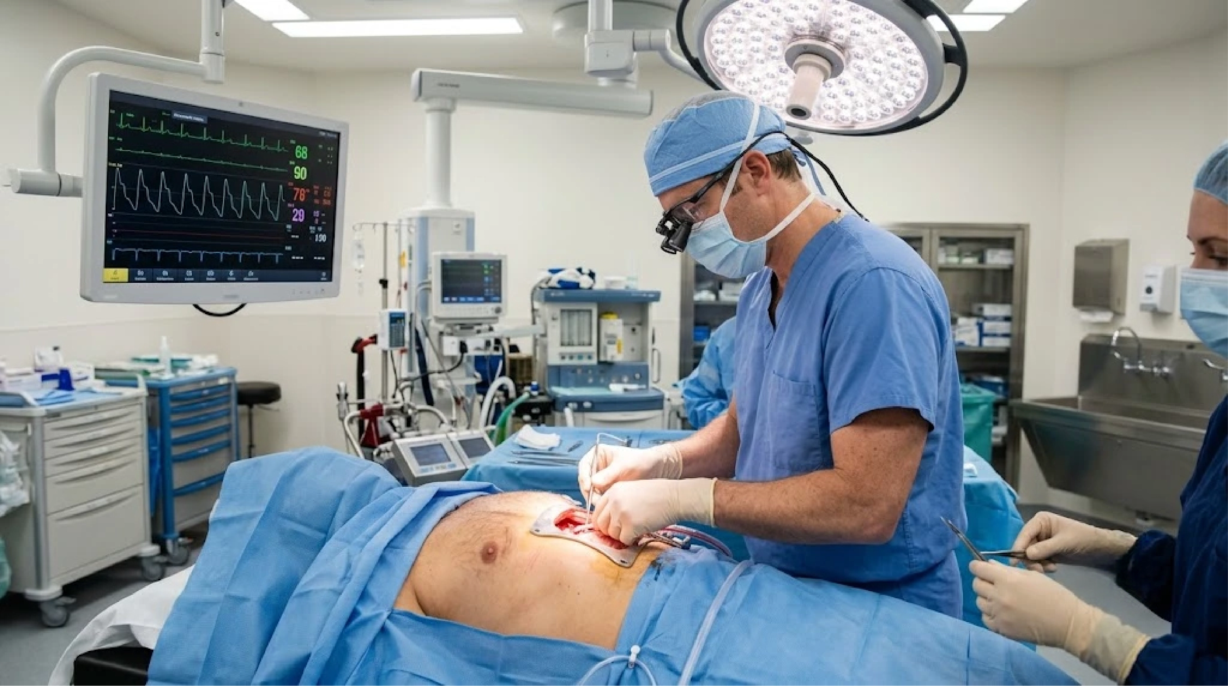
14. Myth: My heart is stopped forever during CABG. Fact: The heart is temporarily arrested using a cold potassium solution to allow the surgeon to sew on a completely still surface. Meanwhile, the heart-lung machine pumps oxygenated blood to the brain and body. The heart is easily and safely restarted after the grafts are complete.
15. Myth: I won't be able to exercise after bypass surgery. Fact: The exact opposite is true. Once the sternum has healed, the restored high-volume blood flow allows patients to exercise with significantly greater capacity, safety, and comfort than they could before their surgery.
Frequently Asked Questions
1. Is an angioplasty painful?
No. You are awake but mildly sedated. Local anesthetic is used at the entry site (usually the radial artery in the wrist, or the groin). You may feel mild pressure or a fleeting ache in the chest when the balloon is inflated, but no sharp surgical pain.
2. How long does a bypass surgery take?
Typically between 3 to 6 hours. The duration depends heavily on the number of bypass grafts required and the complexity of the patient's individual anatomy.
3. Will a heart stent set off airport metal detectors?
No. Coronary stents are microscopic, non-magnetic alloys. They will not trigger airport or security metal detectors.
4. Can I have an MRI scan if I have a heart stent?
Yes. Almost all modern coronary stents are considered MRI-safe immediately or very shortly after placement. Always inform the MRI technician, but having a stent is not a contraindication for an MRI.
5. Where do surgeons get the vessels for a bypass?
The optimal, gold-standard vessel is the Left Internal Mammary Artery (LIMA) located inside the chest wall. Surgeons may also use veins harvested endoscopically from the legs (saphenous vein) or an artery from the forearm (radial artery).
6. Why do cardiac surgeons prioritize the Left Internal Mammary Artery (LIMA) for bypass?
The LIMA is the holy grail of cardiac surgery. Unlike vein grafts, the LIMA is a living, actively functioning artery that secretes high levels of nitric oxide, a powerful vasodilator that naturally repels cholesterol buildup. Because Dr. Sudhir Adalti and our surgical team leave the origin of the LIMA attached to the subclavian artery, it maintains its natural, high-pressure blood flow. This biological property is why over 90% of LIMA grafts function perfectly 15 to 20 years later.
7. How soon after angioplasty can I return to work?
If it was an elective procedure for stable angina, most patients return to desk work within 3 to 7 days. Physical labor will require a slightly longer rest period as dictated by your cardiologist.
8. How soon after CABG can I drive?
Usually 4 to 6 weeks. The breastbone (sternum) needs time to heal. The concussive impact of a deployed airbag or straining heavily on a steering wheel before the bone has fully fused can be dangerous.
9. What is Dual Antiplatelet Therapy (DAPT)?
After a stent is placed, you must take Aspirin plus a second blood thinner (like Clopidogrel, Ticagrelor, or Prasugrel) to prevent blood platelets from sticking to the newly implanted metal mesh while the artery heals over it.
10. How long must I take DAPT?
Typically 6 to 12 months for elective stents, and strictly at least 12 months after a heart attack. Your cardiologist will provide a precise, customized timeline. Never stop these medications without direct physician approval.
11. Is CABG ever done on a beating heart?
Yes. While most CABG is performed using a heart-lung bypass machine (On-Pump), specialized surgeons can perform "Off-Pump" CABG, where a mechanical suction stabilizer holds a small section of the beating heart perfectly still while the delicate graft is sewn.
12. Can blockages return after surgery or stenting?
Yes. Atherosclerosis is an ongoing biological process. If you continue to smoke, have poorly controlled diabetes, or high LDL cholesterol, new plaque can build up in the stents, the bypass grafts, or the native arteries.
13. What is a Drug-Eluting Stent (DES)?
A DES is a metallic stent coated with a microscopic polymer that slowly releases a specialized, localized medication (like everolimus) over several months. This medication prevents excess scar tissue from growing into the stent and blocking it.
14. Are bare-metal stents still used?
Rarely, if ever. Drug-eluting stents have proven to be vastly superior in preventing restenosis and are the undisputed standard of care in advanced cardiovascular centers.
15. What if I have blockages in my leg arteries too?
Atherosclerosis is a systemic disease. If you have coronary artery disease, you may also have Peripheral Arterial Disease (PAD). Comprehensive centers evaluate the entire vascular tree, not just the heart.
16. How does diabetes specifically affect stent success?
Diabetics have a higher biological rate of in-stent restenosis. While modern drug-eluting stents handle diabetes much better than older generations, diffuse diabetic disease is still generally best treated with bypass surgery for long-term safety.
17. When can I resume sexual activity after a heart procedure?
Generally, if you can climb two flights of stairs briskly without chest pain or severe breathlessness, your heart can handle the physical exertion of sexual intercourse. For CABG patients, specific care must be taken regarding physical positions to protect the healing breastbone.
18. Do I need to change my diet after these procedures?
Absolutely. Revascularization fixes the mechanical plumbing, but diet modifies the disease biology. A Mediterranean-style diet, rich in olive oil, fish, and vegetables, and low in saturated fats and refined sugars, is heavily recommended.
19. What happens if a stent gets blocked?
If a stent slowly narrows with scar tissue over years, another balloon or stent can often be placed inside it. If it blocks suddenly with a clot, it causes a heart attack requiring emergency angioplasty.
20. Is the surgical incision from bypass very painful?
Patients are closely managed with multimodal pain medication. Most patients describe a deep ache, soreness, or stiffness in the chest rather than sharp, unbearable pain. The discomfort steadily decreases over the first two weeks of recovery.
Recovery: Adapting to Revascularization
Recovery is not merely about wound healing; it is about how your heart muscle and circulatory system biologically adapt to a sudden influx of highly oxygenated blood after months or years of starvation.
Angioplasty (PCI): Immediate Hemodynamic Relief
- The Physiological Shift: Because PCI involves no general anesthesia or surgical trauma, the heart is not exposed to systemic shock. Once the stent is deployed, ischemic (oxygen-starved) tissue immediately normalizes.
- The Timeline: Patients typically leave the hospital within 24 hours. However, the endothelium (the inner lining of the artery) takes several months to grow over the metallic stent struts-a biological process called endothelialization. During this critical window, strict adherence to Dual Antiplatelet Therapy is mandatory to prevent microscopic clots from forming on the exposed metal.
Bypass Surgery (CABG): Managing "Myocardial Stunning"
- The Physiological Shift: CABG is a systemic event. When the heart is temporarily stopped and placed on a heart-lung machine, the heart muscle undergoes what cardiac intensivists call myocardial stunning. Even after the new grafts are perfectly sewn and blood flow is restored, the heart muscle requires time to "wake up" and regain its full pumping efficiency. Furthermore, the newly harvested internal mammary artery must biologically adapt to the high-pressure demands of supplying the left ventricle.
- The Timeline: The 5 to 7 days spent at Apollo CVHF Hospital are largely dedicated to managing this transition. In our Cardiac ICU, specialists carefully titrate fluids and medications to support the heart while it recovers its contractile strength.
- Sternal Healing: The 6-to-8-week physical restriction on heavy lifting is strictly to allow the sternum (breastbone) to fuse through osteogenesis. Once the bone is fully knit, the patient’s physical capacity is often vastly superior to their pre-surgery baseline.
Cardiac Rehabilitation: The Crucial Next Step
Regardless of whether a patient receives a stent or bypass surgery, participation in a formal Cardiac Rehabilitation program is a critical component of evidence-based medicine. Cardiac rehab is a medically supervised program of clinical exercise training, education on heart-healthy living, and psychological counseling. It has been conclusively proven to reduce hospital readmissions and improve long-term survival.
Why Comprehensive Cardiac Centres Matter
Navigating cardiovascular disease should not be a fragmented journey. Historically, a patient might receive an angiogram at a small clinic, get transferred for surgery to another hospital, and manage their recovery alone at home.
Advanced cardiovascular care requires an integrated ecosystem. Patients with complex heart disease often benefit immensely from the coordinated specialist care available at tertiary referral centers.
A comprehensive cardiac center features:
- Emergency Cardiac Care: A 24/7 catheterization lab capable of halting a STEMI in its tracks with primary PCI within minutes of arrival.
- Advanced Imaging Suites: Access to CT, MRI, and nuclear medicine to phenotype the disease before ever touching the patient.
- Structural Heart Interventions: Expertise in minimally invasive valve replacements (like TAVR) if the patient has co-existing valve disease.
- World-Class Surgical Theaters: Equipped for both complex multi-valve surgeries and minimally invasive bypass procedures.
- Dedicated Cardiac ICUs: Staffed by cardiac intensivists who specialize purely in postoperative heart recovery and hemodynamic monitoring.
- Integrated Preventive Cardiology: To manage cholesterol, hypertension, and rehabilitation seamlessly after the acute procedure is finished.
When these services exist under one roof, the transition from diagnosis to Heart Team discussion, to intervention, and finally to long-term recovery is seamless, incredibly safe, and highly optimized for the patient's long-term survival.
Clinical Insight: The Safety Net of Integrated Care Even with the best planning, a complex angioplasty can occasionally run into complications, such as a severe coronary dissection (tear). In a comprehensive cardiac institute, a cardiothoracic surgical team and operating theater are always on standby in the building. This safety net allows interventional cardiologists to tackle highly complex cases safely, knowing that expert surgical backup is mere steps away.
About Apollo CVHF Hospital
Located in Ahmedabad, Apollo CVHF Hospital is established as a trusted tertiary referral centre for complex cardiovascular care. Functioning as a comprehensive cardiovascular institute, the hospital integrates advanced diagnostic imaging, interventional cardiology, and cardiothoracic surgery to provide multidisciplinary, evidence-based medicine.
The institutional philosophy centers on the Heart Team approach, ensuring that every patient-from those requiring emergency primary PCI to those needing complex, multi-vessel bypass grafting-receives personalized, protocol-driven care. Supported by state-of-the-art intensive care units, structural heart disease expertise, and comprehensive cardiac rehabilitation programs, Apollo CVHF Hospital is dedicated to optimizing long-term patient outcomes through clinical excellence, advanced technology, and collaborative medical expertise.