
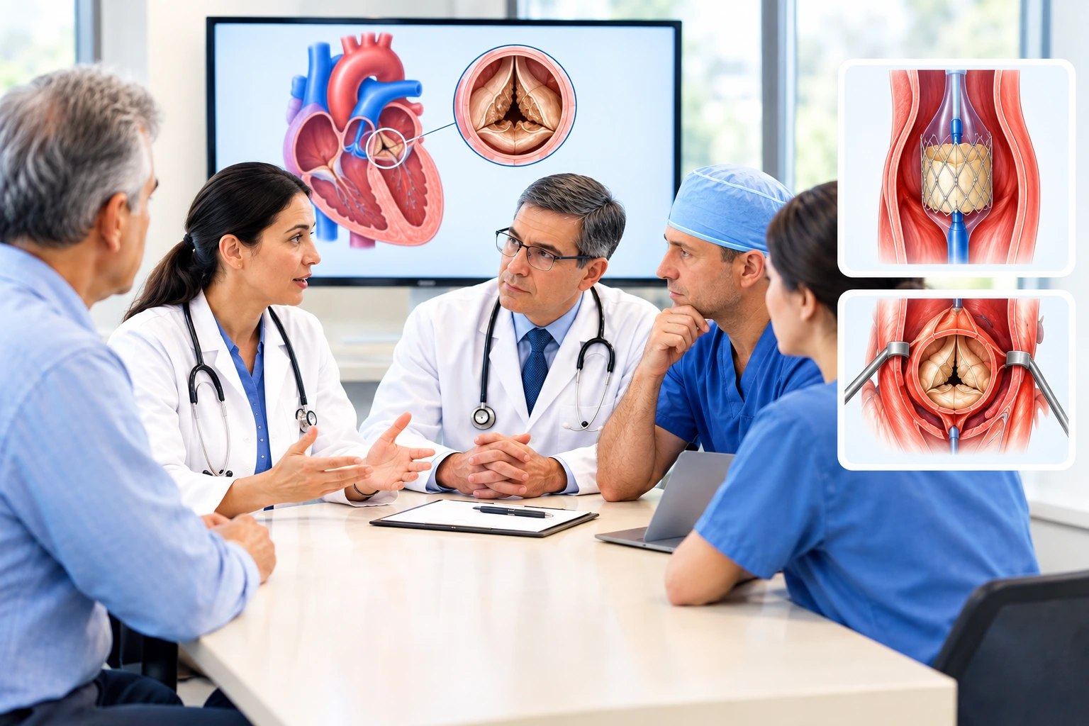
If you or a loved one has been diagnosed with severe aortic stenosis - narrowing of the aortic valve - you're likely trying to understand two treatment paths: TAVI, a catheter-based procedure, and SAVR, traditional open-heart surgery. Both replace the diseased valve. This guide walks through how doctors actually choose between them: confirming the diagnosis, mapping the heart's anatomy in detail, and weighing age, surgical risk, and long-term valve planning together as a team.
Medically reviewed by: Dr. Arvind Singh, Senior Interventional and Structural Cardiologist and Dr. Sudhir Adalti, Senior Cardiothoracic & Vascular Surgeon, Apollo CVHF Hospital, Ahmedabad
Step 1: Confirming Severe, Symptomatic Aortic Stenosis
Aortic stenosis is most often caused by age-related valve calcification. Severity is confirmed by echocardiogram, using measures such as peak jet velocity, mean gradient, and valve area. Once stenosis is severe and causes symptoms - breathlessness, chest discomfort, dizziness, or fainting - outcomes without treatment are poor, and intervention is generally recommended.
Current guidance also supports earlier intervention in carefully selected asymptomatic patients with severe stenosis when procedural risk is low, particularly if there is evidence the left ventricle is starting to be affected (for example, a falling ejection fraction). This is a Heart Team decision, not a default.
Step 2: What Tests Are Usually Needed?
Before either procedure can be planned, a set of tests builds up a full picture of the heart and the patient's overall fitness for treatment:
Echocardiography establishes the severity and haemodynamic consequences of the stenosis (occasionally supplemented by transesophageal echocardiography for more detailed valve imaging); cardiac CT is particularly important for assessing TAVI suitability specifically, and also contributes useful information to surgical planning, though SAVR does not require the same depth of CT-based work-up that TAVI candidacy does.
- Aortic annulus dimensions - to size the replacement valve
- Coronary ostial height - how close the coronary arteries sit to the valve, relevant to the (rare) risk of coronary obstruction during TAVI
- Aortic root anatomy and any aneurysmal disease
- Calcium distribution and burden - asymmetric or bulky calcification affects both TAVI valve seating and surgical planning
- Vascular access - femoral artery size and calcification, since most TAVI is done transfemorally; non-transfemoral access is considered when transfemoral access isn't feasible
- Valve morphology - tricuspid vs. bicuspid, which materially affects the recommendation (below)
|
Test |
What
it tells the Heart Team |
|
Severity of stenosis (jet velocity,
mean gradient, valve area) and left ventricular function |
|
|
ECG |
Baseline heart rhythm and conduction
system, relevant to later pacemaker risk |
|
Valve anatomy, annulus sizing,
coronary height, calcium distribution, and vascular access |
|
|
Whether significant coronary artery
disease is also present |
|
|
Blood tests |
Kidney function, haemoglobin, and
other factors relevant to procedural planning |
|
Clinical assessment |
Symptoms, frailty, comorbidities, and
life expectancy |
Echocardiography establishes the severity and haemodynamic consequences of the stenosis (occasionally supplemented by transesophageal echocardiography for more detailed valve imaging); cardiac CT is particularly important for assessing TAVI suitability specifically, and also contributes useful information to surgical planning, though SAVR does not require the same depth of CT-based work-up that TAVI candidacy does.
The CT scan in particular looks at:
- Aortic annulus dimensions - to size the replacement valve
- Coronary ostial height - how close the coronary arteries sit to the valve, relevant to the (rare) risk of coronary obstruction during TAVI
- Aortic root anatomy and any aneurysmal disease
- Calcium distribution and burden - asymmetric or bulky calcification affects both TAVI valve seating and surgical planning
- Vascular access - femoral artery size and calcification, since most TAVI is done transfemorally; non-transfemoral access is considered when transfemoral access isn't feasible
- Valve morphology - tricuspid vs. bicuspid, which materially affects the recommendation (below)
Step 3: The Heart Team Decision - Current Guideline Framework
Aortic valve treatment decisions are made by a Heart Team: a structural/interventional cardiologist and a cardiac surgeon assessing the same case together, incorporating formal surgical risk scoring (such as STS-PROM or EuroSCORE II) alongside anatomy, age, and life expectancy.
The 2025 ESC/EACTS valvular heart disease guideline sets out the current age- and risk-based framework for tricuspid aortic stenosis:
|
Patient
profile |
Guideline-preferred
approach |
|
?70 years, tricuspid valve, anatomically suitable |
TAVI (Class I), regardless of
surgical risk |
|
<70 years, low surgical risk |
SAVR |
|
All other candidates |
Individualised Heart Team decision,
weighing anatomy, risk, life expectancy, and lifetime valve management |
|
High surgical risk or inoperable, any age |
TAVI |
|
Bicuspid aortic valve |
Anatomy is more complex (asymmetric
calcification, elliptical annulus); TAVI is increasingly used in selected
bicuspid patients with favourable anatomy, but generally carries a
lower-strength recommendation than for tricuspid valves and requires closer
anatomic scrutiny |
This represents a meaningful shift from older frameworks: the age threshold favouring TAVI has moved down from 75 to 70, reflecting accumulating long-term trial data (including low-risk TAVI populations) rather than TAVI being reserved mainly for older or higher-risk patients.
Step 4: Choosing the Valve Type - The Trade-Off That Outlasts the Procedure
Once TAVI or SAVR is chosen as the approach, a separate but related decision is which type of valve to implant - because this affects the patient for years afterward, independent of which procedure was used to place it.
|
Valve
type |
Durability |
Anticoagulation |
|
Bioprosthetic (tissue) valve - used in
TAVI and commonly in SAVR |
Surgical bioprosthetic durability is
well-characterised over decades; TAVI valve durability data is more
established for older patients but still maturing for younger patients now
increasingly receiving TAVI |
Antithrombotic therapy after a tissue
valve depends on the individual - presence of atrial fibrillation, prior
thromboembolism, and other clinical indications for anticoagulation matter as
much as the valve type itself |
|
Mechanical valve - surgical only |
Generally lasts the patient's
lifetime |
Requires lifelong warfarin, with regular
monitoring, regardless of other risk factors |
Why SAVR Remains an Important Option, Not a Fallback
- Concomitant procedures - coronary bypass, another valve repair/replacement, or aortic surgery can be performed in the same operation when needed.
- Mechanical valve option - only available surgically, relevant for younger patients prioritising long-term durability over avoiding anticoagulation.
- Direct surgical access for anatomy that is unfavourable for a transcatheter approach.
Procedure-Specific Risks
TAVI:
- Vascular access complications
- Permanent pacemaker requirement - more common with TAVI than SAVR, due to the valve's proximity to the heart's conduction tissue
- Paravalvular leak
- Stroke
- Rarely, coronary obstruction during valve deployment
SAVR:
- Risks related to cardiopulmonary bypass and general anaesthesia
- Bleeding, infection, and a longer wound-healing period from the sternotomy
- General cardiac surgery risks, including arrhythmia and, rarely, stroke or heart attack
Recovery: Two Different Timelines
- TAVI: many patients mobilise within a day and may be discharged within a few days, though this varies with age, frailty, and whether complications (such as needing a pacemaker) occur.
- SAVR: hospital stay is typically longer, and the sternotomy requires a period of activity restriction (avoiding heavy lifting and pushing/pulling) while the breastbone heals - commonly several weeks, with gradual return to full activity guided by the surgical team.